Dalhousie and Laval students win top prizes at CDA/DENTSPLY event
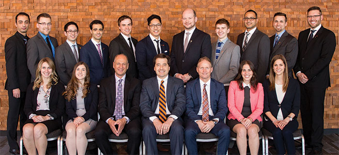
 First prize went to Thomas Steeves of Dalhousie University, for research that compared the effects of 1 versus 3 days of antibiotic treatment on surgical site infections after orthognathic surgery. Based on the results of a double-blind randomized controlled trial, Steeves' study suggests that 3 days of post-operative antibiotics reduced the risk of surgical site infections compared to 1 day, but this benefit may be outweighe
d by the risks of over-administering antibiotics.
"One of our goals in doing this research was to prevent overuse of antibiotics," says Steeves. "We did a number needed to treat analysis that showed only 1 in 10 patients benefitted from the extended 3-day regimen; so this suggests it's still an over-administration of antibiotics. Based on our survey of oral surgeons in Canada, we found that over 40% prescribe oral antibiotics for a week following surgery, so over-prescription of antibiotics may be common."
Steeves won an expense-paid trip to the 2016 American Dental Association (ADA) Annual Meeting in Denver, Colorado, and will present his research at the ADA scientific sessions. "The best part in Vancouver was meeting students from all of the other dental schools," says Steeves. "It was very interesting to compare our clinical experiences and I quickly learned that we are all like-minded."
 Vincent Mireault of Laval University took home second prize. Mireault's research examined the effects of 3 subgroups of unilateral cleft lip and/or palate (cleft lip, cleft lip and palate, and cleft palate) on craniofacial morphology. As runner up, Mr. Mireault received a prize of $1000.
"I was honoured to represent my faculty at this amazing program," says Mireault. "It was very inspiring to meet the other contestants—the future of the profession is surely in good hands!"
The CDA/DENTSPLY Student Clinician Research Program has been running in Canada since 1971 and similar events are held internationally in 36 countries. The program aims to "stimulate ideas, improve communication and, most of all, increase student involvement in the advancement of the dental profession."
A prospective evaluation of 1- versus 3-day post-operative antimicrobial therapy in preventing surgical site infection following orthognathic surgery
By:Thomas Steeves, Dalhousie University (1st Prize)
Advisor: Dr. Curtis Gregoire
Study authors: Thomas Steeves, Dr. Curtis Gregoire, Dr. Clayton Davis
Problem
Orthognathic surgery has a 10-15% risk of surgical site infection (SSI). Pre-operative antibiotic prophylaxis reduces the incidence of SSIs. The recommendations for a post- operative antibiotic prophylactic regimen are conflicting. The purpose of this study is to assess the effect of 3-days versus 1-day of post-operative antibiotic treatment on the prevalence of SSIs following orthognathic surgery.
Methods
A double-blind randomized controlled clinical trial with 288 patients was performed. Inclusion criteria: patients 14 years or older undergoing orthognathic surgery at the Queen Elizabeth II Health Sciences Centre. Exclusion criteria: antibiotic use within two weeks before surgery, and/or active oral or systemic infections. All patients received standard pre-operative antibiotic prophylaxis. Patients were randomly assigned to receive an additional 2-day supplement of either (A) cephalexin (clindamycin for penicillin allergic patients) or (B) placebo. A p-value of <0.05 was considered statistically significant for statistics.
Results & Conclusion
The final cohort had 171 patients after removing those who were non-compliant with antibiotics and those with immunocompromising conditions. Group A and B had 86 and 85 patients, respectively. Three days of antibiotics significantly reduced the prevalence of SSIs compared to 1-day (logistic regression, p=0.04); number needed to treat: 10; odds ratio: 0.34; risk ratio: 0.39. While the extended regimen of antibiotics reduces the risk of SSIs, the benefit may not outweigh the risks of over administering antibiotics and cost ineffectiveness. Compliance may compromise recommendations given to patients.
Comparative analysis of cranio-facial morphology in unilateral clefts: a qualitative report
By: Vincent Mireault, Laval University (2nd Place)
Advisor: Dr. Audrey Bellerive
Objectives
This study aims to compare the specific impacts of the different subgroups of unilateral cleft lip and/or palate (UCLP) - cleft lip (CL), cleft lip and palate (CLP) and cleft palate (CP)- on the cranio-facial development. The dimension and position of the maxilla and the mandible along with the facial height will be the main concerns of this analysis.
Methods
14 CLP, 7 cleft lip (CL) and 16 cleft palate (CP) young patients (mean age of 7 years old) were randomly included in this study. Lateral cephalometric radiographs taken before any orthodontic treatment were analysed in all 37 UCLP patients. One blinded examinator digitally traced all landmarks with the Quick Ceph® Studio program on the radiographs. Standard orthodontic measures were analysed qualitatively to assess the antero-posterior and vertical dimension of both maxilla and mandible as well as their antero-posterior position.
Results
Maxillaries dimension doesn't seem to be affected by UCLP at this developmental stage. However, the maxilla position (SNA) and its inclinaison (NF- (Sn-7)) are different in between UCLP subgroups; CP patients have a more retrusive maxilla whereas CL patients present a more important postero-inferior inclinaison of the maxilla. Most malocclusions observed in all three groups were Cl II Angle and of mild to moderate intensity. CLP seem to have a greater impact on teeth orientation.
Conclusions
Impacts of the different UCLP on the cranio-facial morphology seem to differ slightly in between UCLP subgroups. Major abnormalities—malocclusions—usually observed in CLP might arise later in the development of UCLP patients.
McGill Dental Student Distress and Anxiety: A cross-sectional study from the students' perspective.
By: Erica Abbey, McGill University
Advisor: Dr. Richard Hovey
Study authors: Erica Abbey, Santana Rooyakkers, Karim Rafla, Kristi Stefanison, Dr. Richard Hovey
Problem
Dental students experience high degrees of stress. There is a need to implement stress prevention to not only optimize student mental and physical health but also to enhance learning potential. The purpose of this study is to gain insight and a better understanding of dental students' experience of distress and anxiety during their clinical studies.
Methods
Data was collected from 50 third and fourth year students (25 third year students, 21 males) through a combination of validated surveys; the State-Trait Anxiety Inventory (STAI) and the Distress Thermometer, as well as internally developed surveys.
Results
The STAI scores determined that amongst McGill third and fourth year dental students, 66% had clinically significant symptoms of state anxiety and 62% had clinically significant symptoms of trait anxiety. A majority of students, 85%, rated their distress level above the threshold that has been shown to impede information uptake. There were no reported significant differences between males and females nor third or fourth year students on all assessment tools. Qualitative assessment of student suggestions to reduce distress identified trends relating to scheduling and clinical requirements.
In summary, our study concluded that a significant amount of third and fourth year dental students at McGill University have clinically significant symptoms of anxiety and report high levels of distress. Modifications to the didactic and practical curriculum as well as teaching-learning approaches are suggested to decrease unnecessary distress and demands on students and optimize their health to enhance learning potential.
The new concept of oral cavity diabetes and its significant suppression on cellular prion protein (PrPC) expression: Potential implications in oral cavity development and dental health.
By: Steven A. Arcand, University of Saskatchewan
Advisor: Dr.Changiz Taghibiglou2
Study authors: Steven A. Arcand, Dr.Changiz Taghibiglou2, Brett M. Spenrath1 , Radu I. Stefureac1, Nathan Pham2, Dr. Assem Hedayat1 (faculty contact), Dr.Azita Zerehgar3
1 College of Dentistry, University of Saskatchewan, Saskatoon, Canada
2 Department of Pharmacology, College of Medicine, University of Saskatchewan, Saskatoon, Canada
3 Private Practice
Background
Type II diabetes has a high prevalence in the general and Aboriginal population. Increased caries and gum disease are considered the 7th most common comorbidities in insulin resistant and diabetic patients. We defined a novel concept of 'Oral Diabetes', based on the insulin-signalling pathway in gingival and pulpal tissues of a diet-induced insulin resistance animal model. We also demonstrated that the expression of cellular prion protein (PrPC) in the pulpal tissue of insulin resistant hamsters was suppressed. Since PrPC has been previously identified in oral tissues, the results of this study may explain the high prevalence of periodontal disease and caries in diabetic individuals.
Methodology
Hamsters were fed a high-fructose (n=5) or normal chow (n=5) diet. Upper and lower central incisors, as well as gingival tissues, were excised and biochemically analyzed using ELISA assays and Western blotting for various biomarkers.
Results
Decreased PI3-kinase activity in fructose fed animal models was observed, indicating insulin resistance. A decrease in PIP3 & p-AKT protein levels and an increase in PIP2 levels further confirming this finding. PrPC was detected in pulpal tissues, with a decrease in levels observed in high fructose fed animals.
Conclusion
This is the first reported detection of insulin resistance within the oral cavity. Cellular dysregulation in oral tissues may influence the development of oral pathologies in both the developing and mature dentition. A possible explanation for this may involve altered levels of PrPc expression in insulin resistant oral tissues. These findings may have public health impacts for high-risk populations.
Adhesion-Dependent Interleukin-1 Signaling In Fibroblasts: Role In Periodontal Inflammation
By: Delcorde, J.M., University of Toronto
Advisor: McCulloch, C.A
Study authors: Delcorde, J.M., Wang, Q., McCulloch, C.A.
Problem
Periodontitis is a chronic inflammatory disease characterized by irreversible destruction of alveolar bone and cementum, ultimately resulting in tooth loss. This destruction is mediated in part by the pro-inflammatory cytokine interleukin-1 (IL-1), which signals through adhesive domains (focal adhesions) to enable remodeling of the extracellular matrix. Defining the mechanisms that enable IL-1 signal transduction through focal adhesions could facilitate drug discovery and novel treatments for periodontitis. We hypothesize that sequestration of IL-1 signaling through focal adhesions provides a pivotal regulatory locus, which modulates the temporal kinetics and amplitude of IL-1 signal transduction in human gingival fibroblasts.
Methods
A human transcriptome microarray analysis was performed using gingival fibroblasts with or without intact focal adhesions and IL-1 stimulation. A proteomic analysis of focal adhesion associated proteins was performed on focal adhesion complexes isolated using a pull-down assay employing fibronectin-coated magnetite beads. Samples were labeled with isobaric tandem mass tags and analyzed by liquid chromatography and tandem mass spectrometry for protein identification and relative quantification.
Results
Microarray analysis identified 85 differentially expressed genes induced by IL-1 that also require intact focal adhesions, including kinases and chemokine receptors (fold change >3, p-value < 0.05). Mass spectrometry analysis identified several novel proteins that were recruited to focal adhesions in the presence of IL-1, including Ras suppressor protein 1, serine/threonine-protein kinase PAK2 and Wnt-5a. Understanding the molecular mechanisms that mediate sequestration of the IL-1 signaling pathway to focal adhesions could facilitate the rational design of therapeutics and prognostic markers of inflammatory diseases such as periodontitis.
Regional Developmental Patterns of the Human Mandible
By: Kiavash Hossini, University of British Columbia
Advisor: Dr. Ravindra Shah
Problem
The aims were to use micro-CT scans 1) to analyze 3D growth changes in the mandible during formation of the osseous mandible as it replaces Meckel's cartilage as the main supportive element of the lower jaw; and 2) to compare regional growth patterns of the human mandible during this early development with later periods before and after birth.
Methods
Thirty early fetal specimens (10-19 weeks old; CRL 7.0 -18.1 cm) and eleven late fetal skulls (29-36 weeks old) were scanned with Scanco 100 micro-CT scanner, and another 4 young child skulls (5-6 years old) were scanned using Kodak 90000 CT scanner. The scanned images were viewed and digitized using Dolphin Imaging software and anatomical landmark coordinates were identified for linear and angular 3- Dimensional measurements.
Results
During the early fetal period, the 3D lengths of the primary and secondary mandibular skeletons (Meckel's cartilage to malleus and osseous mandible to condyle) have similar extremely rapid increases in length; most growth (80-90%) occurs in the central body region between the mental foramen and the mandibular lingual. The doubling in length of the body in a few weeks occurs before fusion of the regional osseous components (coronoid, angular and mental), and appears to be supported by growth of Meckel's cartilage and the large carrot-shaped condylar cartilage within the body.
Our results support the concept that abnormal growth of the mandible during this critical early fetal period could have irreversible and long term effects its size and shape.
Evaluation of Nitrile Glove Protection from a Common Dental Contact Allergen
By: Mitchell Naito, University of Alberta
Advisor: Dr. Bernie Linke
Study authors: Mitchell Naito, Dr. Bernie Linke, Daniel Durham, Dr. John F. Elliott
Problem
Methacrylates, a commonly used compound in dentistry, in its uncured form pose a potentially debilitating risk of allergic contact dermatitis to both dental professionals and their patients.
Commonly used dental examination gloves have been previously studied to show methacrylate permeability through the glove, possibly resulting in skin exposure. The objective of our study was to determine glove and skin exposure of methacrylates in a clinical simulation and examine the protection that nitrile gloves provide.
Methods
Samples were obtained in a dental student operative simulation lab by swabbing used nitrile glove surface and the corresponding students skin, after routine student use of composite restoration materials. The swabs were analyzed using gas chromatography-mass spectrometry (GC-MS) for hydroxyethyl methacrylate (HEMA) and triethylene glycol dimethacrylate (TEGDMA).
Results
19 of 22 sampled students had detectable levels of methacrylates on glove surface and 2 students had detectable levels of methacrylates on their skin. A statistical McNemar test showed p values of p<0.01 and p<0.0001 for HEMA and TEGDMA respectively, indicating a statistically significant level of protection provided by the dental gloves.
These results suggest the following: 1) all dental health professionals should wear nitrile when working with methacrylate containing materials, 2) nitrile gloves do not provide 100% protection indicating further material handling precautions, especially for methacrylate-allergic individuals 3) 86% of students had detectable levels of methacrylates on the outer surfaces of the glove, which could potentially serve as a route of exposure to patients.
Patient Experience and Satisfaction of Immediate-Loading: A Qualitative Study
By: Edward Sutton, University of Montreal
Advisor: Dr. Annie St-Georges
Study authors: Edward Sutton, Ryan Luxenberg, Dr. Elham Emami
Objectives
To explore the experience and satisfaction of edentate individuals in regard to the immediate-loading protocol for implant-assisted overdentures.
Methods
Seventeen edentate individuals (mean age: 62 years) who received implant- assisted overdentures through an immediate-loading protocol participated in this qualitative study. An interview guide was developed based on Perneger's Detailed Model and the Health Belief Model. Audio-recorded, semi-structured, in-depth interviews, each with a 60-minute duration, were conducted by two trained interviewers. Qualitative data were analysed using a thematic approach including interview debriefing, transcript coding, data display and interpretation.
Results
Three main themes emerged from the interviews: patient awareness and engagement with treatment, experience-shaped expectations, and immediate gratification. All patients expressed full satisfaction with treatment. Accessibility of the dental care provider and provision of detailed information through proper communication both had a significant impact on patient satisfaction of prosthetic care. High cost and lack of dental insurance coverage were perceived as the major barriers in receiving this type of treatment in the private sector.
Conclusion
The positive experience and satisfaction of patients with the immediate- loading protocol indicates that this modality of treatment should be considered in the treatment planning of edentulous individuals.
Modulation of Myofibroblast Differentiation on Nanometric Topographies
By: Michael T Tiedemann, Western University
Advisor: Dr. Douglas W. Hamilton
Study authors: Michael T Tiedemann, Dr. Douglas W. Hamilton, Dr. Silvia Mittler
Problem
Wound healing is the natural response of tissue to injury in order to re-establish the tissue integrity and structure. Skin and gingival healing comprises of distinct phases; hemostasis, inflammation, proliferation, and remodelling. Recruited fibroblasts in the granulation tissue are responsible for matrix turnover and differentiated myofibroblasts contract the wound edges for closure. In the remodelling phase, inappropriate termination of the healing response can often lead to scarring; excess matrix deposition and persistence of contractile myofibroblasts. Prevention of fibrosis, for example in dental implant aesthetics, could potentially be achieved through patterned scaffold surfaces, but has received relatively little attention. The aim of this study was to compare the effects of surface nanotopographies on human gingival and dermal fibroblast adhesion, differentiation, and extracellular matrix synthesis.
Methods
Human gingival and dermal fibroblasts were seeded on polystyrene slides with nanotopographies patterned on the surface (lines and dots spaced 600, 900, or 1200 nm in size and apart). Immunocytochemistry with fluorescence microscopy was used to assess the fibronectin receptor proteins, β1 and β3, and differentiation of fibroblasts to myofibroblasts with alpha-SMA and the stress fibers of F-actin.
Results
The substratum nanotopography characteristics of surfaces influences cell adhesion through regulation of intracellular signaling events and matrix secretion through integrin receptor-mediated signaling. The nanosurfaces statistically prevent differentiation of fibroblasts into scarring myofibroblasts. Hence, surface topography is a desirable method of modifying cell behavior and permanent method of preventing scarring.
Dentin Preservation in the pericervical area after preflaring with Gates- Glidden or X-Gates drills. An ex vivo micro CT study.
By: Bryan Wong, University of Manitoba
Advisor: Dr. Rodrigo Cunha
Introduction
This study compared the dentin preservation in the distal wall of mesial canals in mandibular molars after preflaring with X-Gates (XG, Dentsply Tulsa Specialities, Tulsa, OK) or Gates-Glidden (GG) drills (Dentsply Tulsa Specialites).
Methods
Sixty mesial canals of mandibular molars were evenly allocated into two balanced groups for preflaring procedures: Group XG – canals were preflared with a single XG drill or Group GG - Drill series (n.1, 2, 3 and 4). With the aid of a micro-computed tomography scanner, dentin removal towards the furcation was measured at 3 levels: furcation (0 mm), 1mm, and 2mm apically. A paired t-test was performed to assess differences before and after procedures in the same group and one-way ANOVA was performed to assess differences between the groups.
Results
Significant decrease in dentin thickness was found in the distal area of mesial roots after preflaring procedures in both groups (P < .0001). No significant difference (P > .05) was observed between XG and GG with regard to dentin preservation. Regarding the level where preflaring was done, significant difference (P < .05) was only detected in the mean values of dentin thickness between 0 mm (411.6 um) and 2mm (251.4 um) in the XG group.
Conclusion
Both XG and GG groups resulted in significant decrease in dentin thickness but no difference was observed between the XG drill group and GG drill group in dentin removal towards the "danger zone."
