


 39 / 48

S
upporting
Y
our
P
ractice
39
Volume 1 Issue 5
|
3
Case Presentation
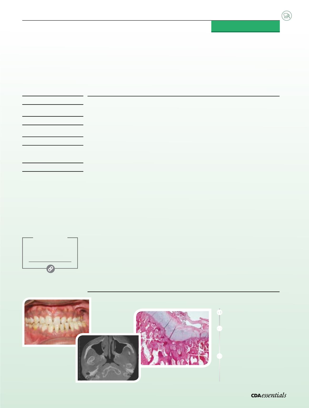
A 41-year-old man was referred to us complaining of jaw dislocation, joint sounds, limitation
of opening and pain on chewing. He reported progressive facial asymmetry that had
developed slowly over 18 months and was clearly visible. Physical examination revealed
mandibular prognathism and a 13-mm deviation of the mandibular midline to the left. The
patient’s maximum jaw opening was 36 mm. A bilateral clicking sound could be heard
during mandibular movements. No pain on palpation of the temporomandibular joints
(TMJs) was present. Masseter muscles were mildly painful to palpation. He had a unilateral
posterior cross-bite on the left side, 4-mm negative horizontal overjet and a class III molar
occlusal relationship (
Fig. 1
).
A panoramic radiograph showed a radiopaque mass attached to the right condyle.
The density of the lesion was similar to that of adjacent bone. The mass had a beak-like
appearance and projected anterior to the right articular eminence. Coronal, axial and
cone-beam computerized tomography images revealed a lesion with cartilaginous features
developing on the condylar head (
Fig. 2
). The lesion had developed medially and superiorly
to pterygoid muscle fibres, causing erosion of the base of the cranial cortex superiorly.
No translation was possible with the right condyle while the left condyle showed a
13.20-mm translation.
An extraoral vertical ramus osteotomy was performed and the proximal segment containing
the condyle and the lesion was removed. The excised tumour measured 3 cm × 2 cm ×
1 cm. Microscopic examination of the decalcified tissue revealed a layer of hyaline cartilage
containing benign chondrocytes in their lacunae. This cartilage formed a cap overlying
normal-appearing trabeculae of cancellous bone (
Fig. 3
). The cartilaginous cap was
covered by a layer of fibrous connective tissue (perichondrium). The osteochondral junction
resembled growth plates with chondrocytes arranged perpendicular to the surface.
What is the diagnosis?
Visit
jcda.ca/article/e16to learn more about the diagnosis and treatment of this case.
a
Severe Unilateral Cross-Bite Secondary to
Tumour of theMandibular Condyle
The following is a condensed version of an article published in the
‘Clinical Dentistry’ section of
jcda.ca—CDA’s online, open access
scholarly publication that features articles indexed in Medline,
Journal Citation Reports and Science Citation Index.
Diagnostic Challenge
Nathalie Rei
DMD,MSc
Normand Bach
DMD,MSc, FRDC(C)
Michel El-Hakim
DMD,MD,MSc,
FRDC(C), Dip ABOMS
Adel Kauzman
DMD,MSc, FRDC(C)
➊
Frontal view
of the occlusion at
presentation.
➋
Axial cone-beam
computerized tomography
shows a mass anterior and
medial to the condylar head.
➌
Photomicrograph of the
decalcified specimen
showing
a hyaline cartilage cap covering
cancellous bone trabeculae
and fatty marrow.
More Online
Access the full-text
article at:
jcda.ca/article/e16➌
➊
➋