|
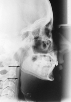
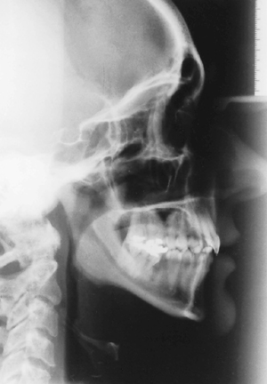
| Figure 1a: Lateral cephalometric radiograph of a patient with obstructive sleep apnea syndrome (OSAS) indicates the narrow anteroposterior dimension of the pharyngeal airway at the level of the soft palate and the base of tongue (arrows). |
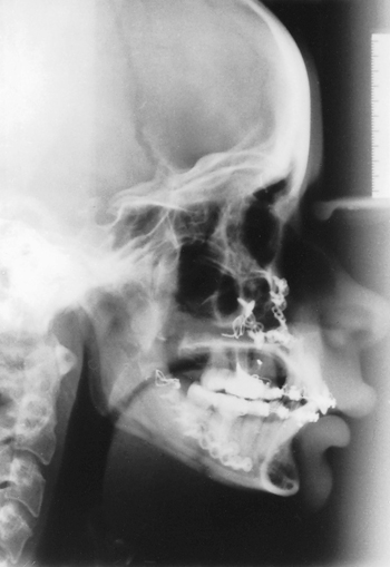 |
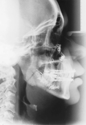
| Figure 1b: Postoperative lateral cephalometric radiograph of the same patient after maxillo- mandibular advancement. Note the signicant increase in the pharyngeal airway space at the level of the soft palate and the base of tongue. |
 |
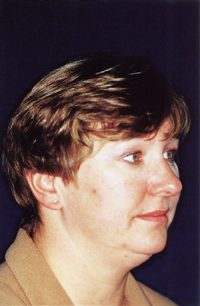
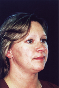
| Figure 2: Preoperative photograph of the patient depicted in Figure 1.Patients without a clinically obvious facial deformity may benefit from maxillomandibular advancement. As this photograph indicates, patients with OSAS may not be obese. |
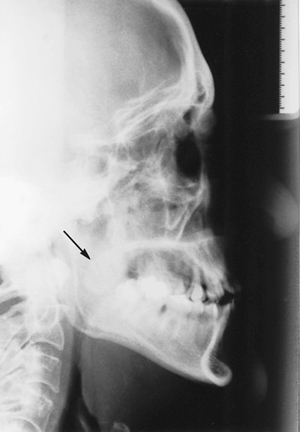 |
| Figure 3: Lateral cephalometric radiograph of a patient with OSAS who underwent uvulopalato- pharyngoplasty demonstrates a thick, bulbous soft palate (arrow) associated with a narrow naso- pharyngeal airway. |
 |
| Figure 4a: Preoperative photograph of a patient with OSAS. |
 |
| Figure 4b: Postoperative photograph of the same patient 6 months after maxillomandibular advancement. |
 |
| Figure 4c: Preoperative lateral cephalometric radiograph of the same |
 |
| Figure 4d: Lateral cephalometric radiograph of the same patient after maxillomandibular advancement shows signi cant improvement in the pharyngeal airway space. |