|
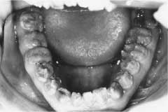
| Figure 1: Hypoplastic amelogenesis imperfecta. Clinical photograph shows irregular enamel formation and severe attrition. |
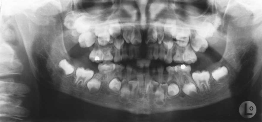 |
| Figure 2: Cleidocranial dysplasia. Panoramic radiograph shows multiple supernumerary teeth and retained deciduous teeth. |
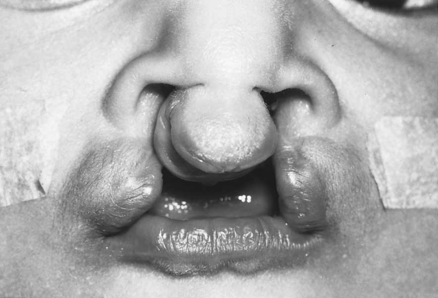 |
| Figure 3: Bilateral cleft lip and cleft palate. |
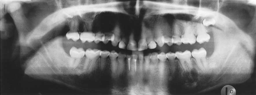 |
| Figure 4: Dentinogenesis imperfecta. Panoramic radiograph shows obliteration of pulp and periapical abscess of tooth 36. |
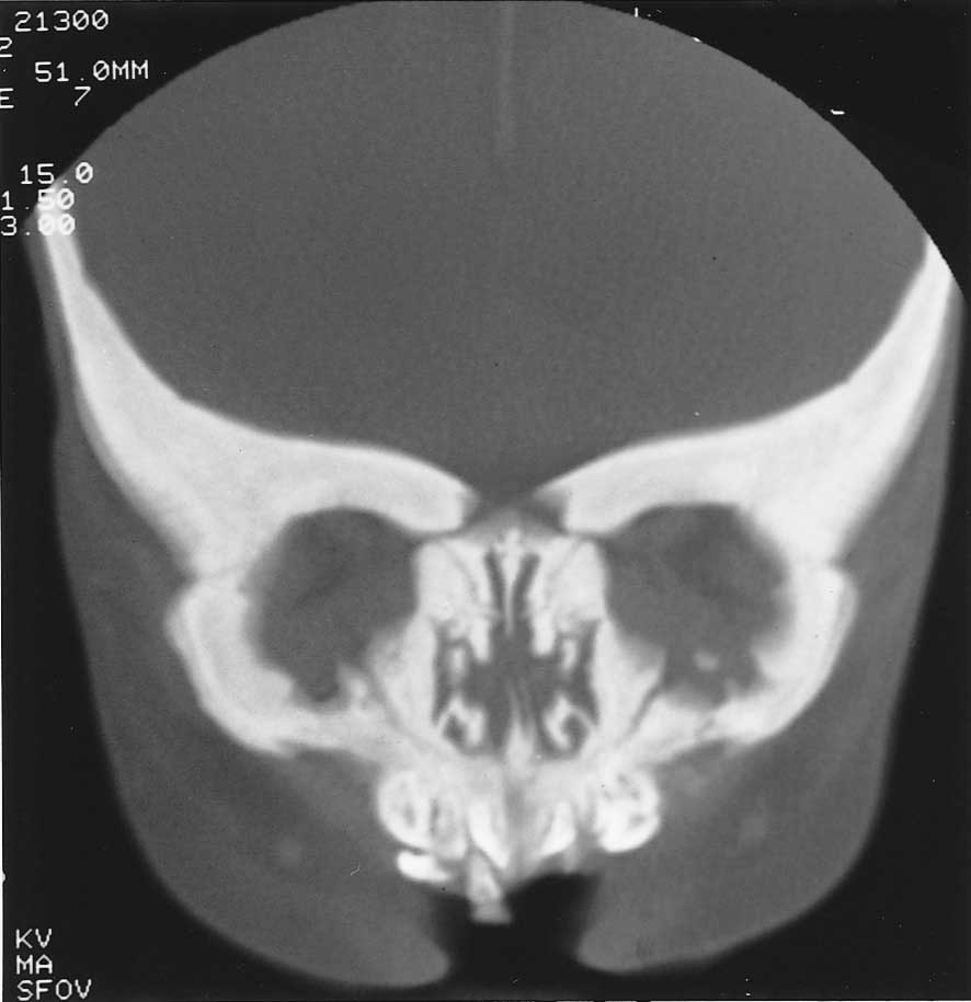 |
| Figure 5: Osteopetrosis. Computed tomography (bone window) of the head and neck shows increased density of bone, obliteration of marrow spaces and increased thickness of cortical bone. |
 |
| Figure 6: Mandibulofacial dysostosis. |
 |
| Figure 7: Oligodontia, imaged with panoramic radiography. |
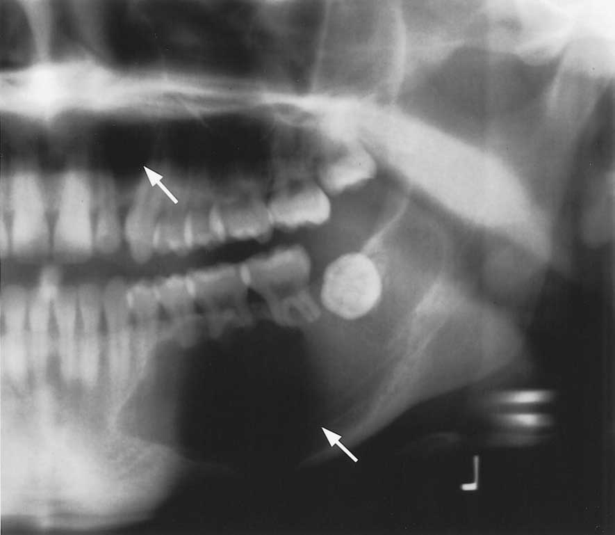 |
| Figure 8: Nevoid basal cell carcinoma syndrome. Panoramic radiograph shows multiple jaw cysts (white arrows), in particular one in the maxilla between teeth 22 and 23 and another in the body and ascending ramus of the mandible, causing downward displacement of the inferior alveolar nerve and resorption of the roots of permanent teeth 34, 35, 36 and 37. The third permanent molar (tooth 38) is transversely impacted. |
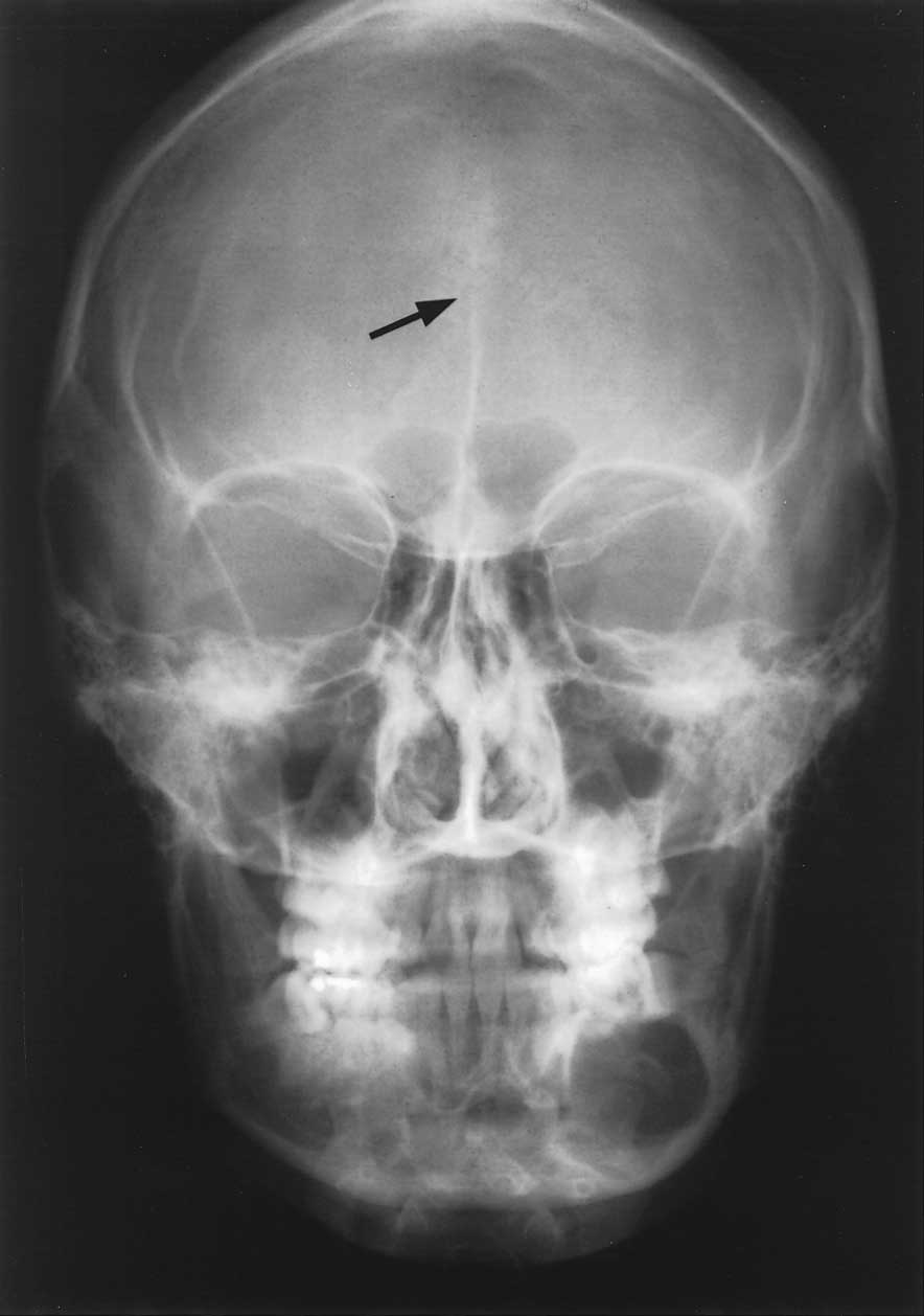 |
| Figure 9: Nevoid basal cell carcinoma syndrome. Posteroanterior cephalogram shows calcification of the falx cerebri (black arrow). |