|
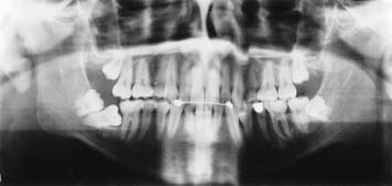
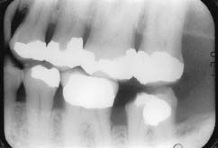
| Figure 1: Panoramic radiograph revealing horizontally impacted teeth 47 and 48. Note the stage of root formation of tooth 48. |
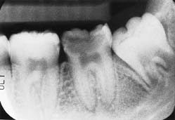 |
| Figure 2: Periapical radiograph of the left posterior mandible demonstrating extensive decay associated with tooth 37. Note the stage of root development of tooth 38. |
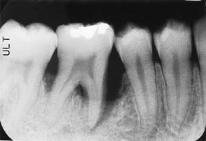 |
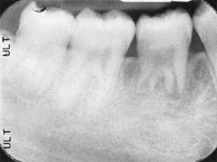
| Figure 3: Periapical radiograph showing localized juvenile periodontitis associated with tooth 46. |
 |
|
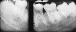
(a)
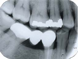
(b) Figure 4: Periapical radiographs of the lower third molars show that root development of tooth 48 (a) appears greater than two-thirds; therefore, tooth 38 (b) was used as the donor tooth. |
 |
| Figure 5: Six-month post-operative radiograph indicates patient has regained the supporting alveolar bone in the region of the tooth transplant and shows continued root development with the establishment of a periodontal ligament space. |
 |
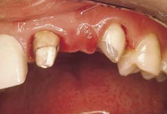
| Figure 1: Dislodged anterior porcelain-fused-to-metal fixed partial denture one year after initial insertion. The prosthesis is in excellent condition, but the supporting structure had design errors. |
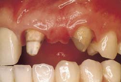 |
| Figure 2: The abutment teeth supporting the fixed partial denture shown in Fig. 1. Note the conical shape of the canine abutment and the excessive angle of convergence of the incisor abutment. |
 |
| Figure 3: Following cleaning, the surfaces of the abutment teeth were etched with a phosphoric acid etchant gel for 20 seconds. |
 |
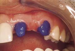
| Figure 4: Following etching of the abutment teeth, the surfaces were primed with a dentin bonding agent. Note the shiny appearance of the dentin surfaces after this step. The shine is a good indication of thorough priming. |
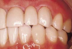 |
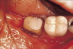
| Figure 5: Post-operative view after cementation of the original prosthesis with a resin cement. Note the excellent colour match. |
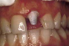 |
| Figure 6: This maxillary central incisor had an all-porcelain crown that dislodged just one year after initial insertion. The tooth was prepared with an excessive angle of convergence. |
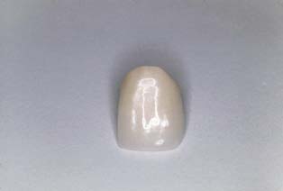 |
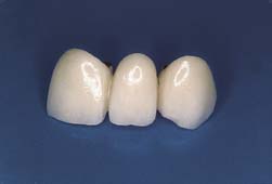
| Figure 7: This all-porcelain crown belonged to the tooth in Fig. 6. Note the excellent condition of the crown. |
 |
| Figure 8: Bitewing radiographic image of mandibular second molar tooth from which a crown dislodged. |
 |
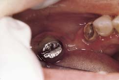
| Figure 9: The mandibular second molar shown in Fig. 8 after restoration with a resin composite material and extending the original crown preparation subgingivally. |
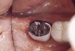 |
| Figure 10: The fabricated porcelain-fused-to-metal crown was cemented with a resin cement following a bonding procedure to enhance retention. The occlusal surface of the crown had to be made in metal to minimize occlusal reduction and thus maximize crown height. |
 |
| Figure 11: A posterior porcelain-fused-to metal fixed partial denture dislodged in this case due to recurrent caries in the mesial abutment. |
 |
| Figure 12: Radiographic image of the fixed partial denture just over 7 years after initial recementation with a dentin-bonded resin cement. The recementation helped to maintain the integrity of the dental arch over the years and preserved the abutment teeth from caries. |
Table 1