

![]()
![]()
Midfacial Complications of Prolonged Cocaine Snort
Peter D. Villa, DDS, FRCD(C)
ABSTRACT
Acute and chronic ingestion of cocaine predisposes the abuser to a wide range of local and
systemic complications. This article describes the case of a 38-year-old man whose chronic
cocaine snorting resulted in the erosion of the midfacial anatomy and recurrent sinus
infections. Previously published case reports specific to this problem are presented, as
are the oral, systemic and behavioural effects of cocaine abuse.
MeSH Key Words:Words: case report; cocaine; substance-related disorders.
© J Can Dent Assoc 1999; 65:218-23
This article has been peer reviewed.
[Introduction| Case Report | Pharmacology |Clinical Findings of Cocaine Abuse |Literature Review |Summary |References]
It is estimated that two million Americans are addicted to cocaine.1 In Ontario, a survey by the Addiction Research Foundation found that almost 5% of the adult population had used cocaine at least once in their lifetime.2 Much of the recent literature on this subject has focused on the behavioural and systemic effects of cocaine abuse as well as on drug interaction considerations for the management of dental patients who are addicts.3-9 This article describes the devastating midfacial deterioration suffered by a cocaine snorter. A brief overview of the clinical dental findings is provided and considerations for the management of patients with cocaine abuse problems are discussed.
On February 3, 1998, a 38-year-old man was seen for evaluation of an oral-nasal communication after having been referred by his family dentist. The patient described how problems began to manifest themselves as nosebleeds in July 1997 and how, during the following months, those symptoms progressed to recurrent sinus infections. He first noticed a "pinhole" in his palate in late November 1997, after a soft drink he consumed ran out his nose. The opening became larger over the next two months, stabilizing in size to the diameter of his little finger. The patient discovered that a thick layer of bubble gum could be used to cover the defect, normalize his speech, and prevent food stuffs from being displaced into his nose.
The patient’s medical history indicated years of repeated cocaine snorting and a smoking habit of one-half pack of cigarettes per day. He was employed as a labourer, renovating the interior of commercial buildings.
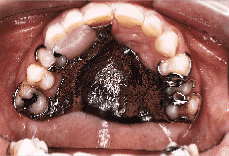
The patient displayed a saddlenose deformity, characterized by a broad, flat nose (Fig. 1). There was no facial swelling, cervical lymphadenopathy, intraoral swelling, or trismus. Primary tooth 53 was deeply decayed and permanent cuspid tooth 13 was erupting palatally. A 10 x 12 mm oval fistula was apparent through the roof of his palate, just left of the midline, in the first molar area. No drainage or exophytic lesions were apparent.
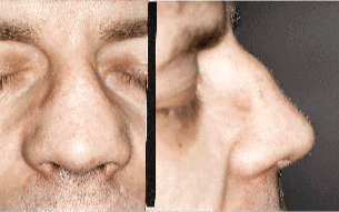 |
| Fig. 1: Saddlenose deformity, front and side views. |
Midline Lethal Granuloma, Wegener’s Granulomatosis, nasal lymphoma, and tertiary syphilis can all present with these clinical findings.10-12 The patient’s workup therefore included a biopsy of the palatal mucosa, computed tomography (CT) scans, ear, nose and throat (ENT) evaluation, complete blood count (CBC), sedimentation rate, antinuclear antibody test (ANA), venereal disease test (VDRL), chest x-ray, and urinalysis. After consultation with specialists in other disciplines, results of these tests increased our confidence that we were dealing only with the local effects of cocaine abuse. Figure 2a is a CT scan of the patient’s nasopalatal defect, while Fig. 2b shows a CT scan of a normal midfacial anatomy.
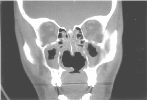 |
| Fig. 2a: CT scan showing palatal perforation, loss of nasal septum and turbinates, and thickening of the maxillary sinus membranes. |
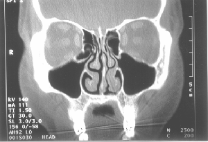 |
| Fig. 2b: CT scan of normal midfacial anatomy. |
The biopsy of soft tissue, taken from the palatal margin of the oral-nasal opening, revealed a non-specific ulcer and chronic inflammation with some eosinophils. The presence of eosinophils has been noted in pathologists’ findings, as reported in Armstrong and Shikani10 and Schweitzer.13
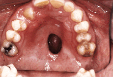
Management was predicated on complete cessation of the drug. The patient was informed of the consequences of continued cocaine use, and how to get help in quitting. He was also advised to smoke less, and to use a proper filtration mask while at work. Appropriate management of recurrent sinus infections was coordinated with his family physician. After basic oral hygiene and restorative procedures were provided, a removable obturator was constructed (Fig. 3a, 3b and 3c). The patient will be re-evaluated for possible surgical closure of the oral-nasal fistula at a later date.
 |
 |
| Fig. 3a: Nasopalatal defect. | Fig. 3b: Obturator removed |
 |
| Fig. 3c: Obturator inserted. |
Pharmacology
Cocaine is a naturally occurring alkaloid. It is extracted from the leaves of the Erythroxylon coca plant, which is indigenous to three countries in northern South America.4 Cocaine is a psychologically disruptive and dependence-inducing drug; classified as a psychostimulant, it exhibits both local anesthetic and neurotransmitter effects.5,11,13 Like lidocaine, it functions as a local anesthetic by blocking the sodium channels of neural tissues, and like lidocaine, can trigger seizures at higher doses.14 Its neurotransmitter effects are attributed to a blocking action on the reuptake of specific transmitter agents by the presynaptic nerve endings. The resultant excess of neurotransmitter causes increased stimulation of the postsynaptic nerves. Dopamine activity is enhanced in the brain, causing a feeling of euphoria.15 Peripherally, norepinephrine is the transmitter whose activity is increased.11 This profound enhancement of sympathetic tone is responsible for the vasoconstrictive, tachycardiac, and dysrhythmic actions of the drug.6,8,14,16,17
Cocaine also affects pulmonary physiology. By acting at the level of the medulla, an increase of the respiratory rate is produced.4 It has been postulated that vasoconstriction of the pulmonary circulation reduces blood flow sufficient to induce hypoxia.4 This is significant when one considers that the cardiovascular effects of cocaine profoundly increase myocardial oxygen demand while simultaneously vasoconstricting the coronary arteries.5 The potential then exists for myocardial infarction, pulmonary edema, circulatory collapse, and death.6,16
Cocaine is well absorbed from mucous membranes and the gastrointestinal mucosa. It is rapidly degraded by hepatic and plasma esterases to water soluble metabolites that are excreted in the urine.5,16 Peak blood levels occur within 30 minutes, with most of the drug gone within two hours.18 While trace amounts of cocaine may be found in the bloodstream for eight to 12 hours after drug use, metabolites may be present for ten days.5
Cocaine is commonly taken intravenously, by smoking or inhalation of the "crack" or "freebase" form, or by snorting.5,8,13 Although less common, cocaine can also be topically applied to gingival tissues, or ingested orally (mixed with cocktails).13,19,20 Cocaine has an acidic pH of 4.0; it’s purity and sterility, and the type of adulterants it is mixed with, all directly affect its potential for local and systemic complications.17,21,22 HIV, hepatitis, and endocarditis are more prevalent in the population of intravenous drug abusers.3,5,7,13,16
Clinical Findings of Cocaine Abuse
The street form of cocaine is both vasoconstricting and locally irritating to the thin respiratory epithelium of the nasal airway. Repeated snorting sets up a cascade of ischemia, inflammation, micronecrosis, infection, and then macronecrosis leading to perforation.11,23 Nasal septum perforations of both the cartilaginous and bony tissues have been well documented.3,24 With larger defects, support of the nose is compromised, resulting in the typical saddlenose deformity.3,24 Some patients have been known to use various narrow instruments to debride intranasal crusting, increasing the potential for perforations.11 In extreme cases, adjacent bony structures may become eroded and vital tissues damaged.6,12,13,22,23
Similarly, topically applied cocaine can be locally destructive to the oral mucosa and dentition. Acute ulceration, necrosis, and rapid recession of gingival tissues, as well as erosion of both dentin and enamel, have been reported.19,20 Inhalation of "crack" cocaine has been implicated in the corrosion of gold dental restorations.25 Moreover, cocaine consumption immediately before or after tooth extraction can result in excessive hemorrhage.26
Several publications list other oral findings that are indirectly associated with cocaine abuse.4,7,20,25 Patients with a substance abuse problem will frequently display higher rates of decay and periodontal disease as a result of general neglect.4,7,25 Chronic cocaine users often develop bruxing habits and demonstrate patterns of severe occlusal wear.4,7,20 Aggressive tooth brushing while on a "cocaine high" has been implicated as the cause of both cervical tooth abrasion and gingival lacerations.4,7 Xerostomia and oral candida infections are also more common in this patient population.4,7,25
Literature Review
The case of a 37-year-old woman who developed a palatal defect several years after a nasal septal perforation is described by Sastry and others.11 Her long history of cocaine abuse continued despite initial violation of the septal structure. The authors postulate that vigorous self-debridement of intranasal crusts with cotton swabs, pens, and pencils contributed to the perforation process. Unfortunately, such debridement is well tolerated because of the profound local anesthetic effects of cocaine.
In another case, Sawicka and Trosser detail the findings of a 34-year-old man who presented himself at the hospital with a six-day history of clear nasal discharge and malaise.23 The patient, who had lost his sense of smell, admitted to a 19-year habit of cocaine snorting. A CT scan showed bone loss of the cribriform plate, and suggested a cerebrospinal fluid (CSF) leak through the right ethmoid sinus. A bifrontal craniotomy and fascia lata graft were performed to correct the persistent leak. The cribiform plate was noted to be paper thin and mobile. Histology of the olfactory bulb showed chronic inflammation change and gliosis.23
Cocaine abuse can cause other complications. Newman and others report the case of a 43-year-old man with bilateral optic neuropathy and osteolytic sinusitis, secondary to cocaine abuse.22 The patient had initially described "holes" in his vision that progressed over a six-month period. He admitted to a 15-year history of daily intranasal cocaine use. MRI studies revealed extensive bony destruction of the nasal cavity, paranasal sinuses, the floor of the anterior cranial fossa, and the anterior surface of the clivus. After a four-month cessation of cocaine use, his visual acuity stabilized and his visual field deficits had not progressed.22
Schweitzer describes two patients with severe and different complications as a result of cocaine abuse.13 The first patient developed total nasal septal necrosis, saddlenose deformity, and osteolytic sinusitis from chronic snorting. Her presenting symptoms included a five-year history of postnasal drainage, halitosis, intermittent epistaxis, and rhinitis. After a proper workup and detoxification, the patient underwent bilateral antrostomies and nasal reconstruction with auricular cartilage. With daily saline lavages of the nose and sinuses, her perinasal symptoms subsided. The second patient experienced tracheobronchial rupture with subcutaneous emphysema and pneumomediastinum after smoking "freebase" cocaine.
One of the most destructive cases of intranasal cocaine abuse to have been documented appears in the journal Revista Medica de Panama, where Sousa and Rowley detail the presenting complications, progression, and eventual death of a 22-year-old woman.12 In this case, the patient described a two-year history of nasal obstruction, halitosis, progressive destruction of the septum and hard palate, purulent rhinorrhea, intense facial pain, strabismus, blindness in her left eye, and a recent reduction in the visual acuity in her right eye. Her diagnostic workup included physical, ophthalmoscopic, and rhinoscopic examinations, multiple biopsies, bacterial and fungal cultures, and CT scans. These studies confirmed the absence of the nasal septum, turbinates, and medial walls of the maxillary sinuses. They also revealed sclerosis at the base of the skull and a midline lesion extending from the ethmoid sinuses to the orbital apexes. Initial treatment with prednisone and antibiotics resulted in improvement of the visual acuity in her right eye and resolution of the retro-ocular pain. Several months later, suspected of having renewed her drug habit, the patient was readmitted to hospital with meningitis. Her level of consciousness began to deteriorate on the twelfth day. A brain scan revealed an abscess within her frontal lobe. An emergency craniotomy was performed. The patient remained comatose and on a ventilator for 15 days. Death occurred as a result of Pseudomonas pneumonia.
Other cases of brain abscesses resulting from habitual cocaine snorting have been reported.21,27 Possible routes of bacterial inoculation include direct spread through the areas of osteitis (i.e. cribriform plate, frontal sinus) or as a septic thrombophlebitis spread along the associated valveless venous vasculature.21 These expanding cerebral abscesses are usually fatal.12,21
Recreational drug use is reaching epidemic levels in North America. There are numerous considerations in the provision of dental care for patients with a cocaine abuse problem. Given the fundamental importance of identifying whether cocaine is a factor in the patient’s management, the dentist should look for signs and symptoms indicating an abuse problem (Tables I and II). An appropriate medical history, a detailed examination of the orofacial anatomy, routine vital signs, and an understanding of the behavioural characteristics of an addict will help the practitioner recognize patients suspected of cocaine abuse. A patient with a substance abuse problem will frequently exhibit "drug-seeking" behaviour.
| Table I | |
| Orofacial Signs and Symptoms of Chronic Cocaine Abuse | |
| Snorting | Gingival application |
| Table II | |
| Effects of Cocaine Abuse | |
| Cardiovascular effects | Central and behavioural effects |
The family dentist should know that the injection of local anesthetic with epinephrine must be avoided for at least six hours after cocaine consumption.18 Some sources suggest the use of epinephrine in either local anesthetic or retraction cord is contraindicated for at least 24 hours after cocaine use to prevent "sympathetic overload" resulting in a hypertensive crisis, cerebrovascular bleed, myocardial infarction, tachydysrhythmias, and/or cardiac arrest.21,28 Lidocaine without vasoconstrictors will have an additive effect with existing cocaine in reducing the patient’s threshold for seizure activity.4,5 As well, general anesthesia poses significant cardiovascular risk and should be avoided with the chronic cocaine user.4
Ingesting powdered cocaine orally or nasally can be extremely destructive to the periodontal and midfacial anatomy. Once alerted to an abuse problem, the informed dentist can educate his or her patient about the progressive consequences of continued usage and provide a referral for professional counselling. Dental treatment should be deferred to an appropriate time when life-threatening complications can be avoided. Then, successful restorative, periodontal, and even obturator therapy can be provided.
An understanding of and vigilance for cocaine abuse in the dental patient can reduce, but will not eliminate, the potential for a related crisis in the dental office. Dental practitioners and their staff should remain capable of recognizing and managing a cocaine-related medical emergency. Dentists and dental societies must continue to educate the general public about the local and systemic hazards of this drug.
Dr. Villa is an oral and maxillofacial surgeon and head of dentistry at The Sudbury Regional Hospital in Ontario. He also maintains a private practice in Sudbury.
Acknowledgments: A special thanks to Olivia Simonetti for her translation services in the preparation of this article and to Dr. Michael Hamilton for his services in the care of the patient described in this case report.
Reprint requests to: Dr. Peter D. Villa, 306-2009 Long Lake Rd., Sudbury, ON P3E 6C1
References
1. Gawin FH. Cocaine abuse and addiction. J Fam Pract 1989; 29:193-7.
2. Adlaf EM, Ivis F, Walsh G, and Bondy S. Alcohol, tobacco, and illicit drug use amongst Ontario adults. 1977-1996. Survey by the Addiction Research Foundation.
3. Laskin DM. Looking out for the cocaine abuser. J Oral Maxillofac Surg 1993; 51:111.
4. Lee CY, Mohammadi H, and Dixon RA. Medical and dental implications of cocaine abuse. J Oral Maxillofac Surg 1991; 49:290-3.
5. Goldstein FJ. Toxicity of cocaine. Compendium 1990; 11:710, 712, 714-6.
6. Pallasch TJ, McCarthy FM, and Jastak JT. Cocaine and sudden cardiac death. J Oral Maxillofac Surg 1989; 47:1188-91.
7. Friedlander AH, Gorelick DA. Dental management of the cocaine addict. Oral Surg Oral Med Oral Pathol 1988; 65:45-8.
8. Isaacs SO, Martin P, and Willoughby JH. "Crack" (an extra potent form of cocaine) abuse: a problem of the eighties. Oral Surg Oral Med Oral Pathol 1987; 63:12-6.
9. Ravi VS, Zmyslowski WP, and Marino J. Probable cocaine-induced hyperthermia in an anesthetized patient: a case report. J Oral Maxillofac Surg 1993; 51:204-5.
10. Armstrong M Jr, Shikani AH. Nasal septal necrosis mimicking Wegener’s granulomatosis in a cocaine abuser. Ear Nose Throat J 1996; 75:623-6.
11. Sastry RC, Lee D, Har-El G. Palatal perforation from cocaine abuse. Otolaryngol Head Neck Surg 1997; 116:565-6.
12. Sousa O, Rowley S. [Otorhinolarygologic symptoms caused by the intranasal abuse of cocaine. Report of a case.] Rev Med Panama 1994; 19:55-60.
13. Schweitzer V. Osteolytic sinusitis and pneumomediastinum: deceptive otolarygologic complications of cocaine abuse. Laryngoscope 1986; 96:206-10.
14. Isner JM, Estes NA 3d, Thompson PD, Castanzo-Nordin MR, Subramanian R, Miller G, and others. Acute cardiac events temporally related to cocaine abuse. N Engl J Med 1986; 315:1438-43.
15. Woolverton WL, Johnson KM. Neurobiology of cocaine abuse. Trends Pharmacol Sci 1992; 13:
193-200.
16. Das G. Cardiovascular effects of cocaine abuse. Int J Clin Pharmacol Ther Toxicol 1993; 31:521-8.
17. Estroff TW, Gold MS. Medical and psychiatric complications of cocaine abuse with possible points of pharmacologic treatment. Adv Alcohol Subst Abuse 1985; 5:61-76.
18. Little JW, Falace DA. Dental management of the medically compromised patient. 3rd ed. St. Louis (MO): The C.V. Mosby Co; 1988.
19. Kapila YL, Kashani H. Cocaine-associated rapid gingival recession and dental erosion. A case report. J Periodontol 1997; 68:485-8.
20. Parry J, Porter S, Scully C, Flint S, Parry MG. Mucosal lesions due to oral cocaine use. Br Dent J 1996; 180:462-4.
21. Rao AN. Brain abscess: a complication of cocaine inhalation. NY State J Med 1988; 548-50.
22. Newman NM, DiLoreto DA, Ho JT, Klein JC, Birnbaum NS. Bilateral optic neuropathy and osteolytic sinusitis. Complications of cocaine abuse. JAMA 1988; 259:72-4.
23. Sawicka EH, Trosser A. Cerebrospinal fluid rhinorrhoea after cocaine sniffing. Br Med J (Clin Res Ed) 1983; 286:1476-7.
24. Meyer R. Nasal septal perforations must and can be closed. Aesthetic Plast Surg 1994; 18:345-55.
25. Brown RS, Johnson CD. Corrosion of gold restorations from inhalation of "crack" cocaine. Gen Dent 1994; 242-6.
26. Johnson CD, Brown RS. How cocaine abuse affects post-extraction bleeding. JADA 1993; 124:60-2.
27. Brown E, Prager J, Lee HY, Ramsey RG. CNS complications of cocaine abuse: prevalence, pathophysiology, and neuroradiology. AJR Am J Roentgenol 1992; 159:137-47.
28. Goulet JP, Perusse R, Turcotte JY. Contraindications to vasoconstrictors in dentistry. Part III: Pharmacology interactions. Oral Surg Oral Med Oral Pathol 1992; 74:692-7.